Neuronal and oligodendroglial, but not astroglial, tau translates to in vivo tau PET signals in individuals with primary tauopathies
2024.11.24.
Luna Slemann et al.
Acta Neuropathol, 2024
Summary
Tau PET has attracted increasing interest as an imaging biomarker for 4-repeat (4R)-tauopathy progressive supranuclear palsy (PSP). However, the translation of in vitro 4R-tau binding to in vivo tau PET signals is still unclear. Therefore, we performed a translational study using a broad spectrum of advanced methodologies to investigate the sources of [18F]PI-2620 tau PET signals in individuals with 4R-tauopathies, including a pilot PET autopsy study in patients. First, we conducted a longitudinal [18F]PI-2620 PET/MRI study in a 4-repeat-tau mouse model (PS19) and detected elevated [18F]PI-2620 PET signals in the presence of high levels of neuronal tau. An innovative approach involving cell sorting after radiotracer injection in vivo revealed higher tracer uptake in single neurons than in the astrocytes of PS19 mice. Regional [18F]PI-2620 tau PET signals during the lifetime correlated with the abundance of fibrillary tau and with autoradiography signal intensity in PSP patients and disease controls who underwent autopsy 2–63 months after tau PET. In autoradiography, tau-positive neurons and oligodendrocytes with a high AT8 density, but not tau-positive astrocytes, were the drivers of [18F]PI-2620 autoradiography signals in individuals with PSP. The high tau abundance in oligodendrocytes at the boundary of gray and white matter facilitated the identification of an optimized frontal lobe target region to detect the tau burden in patients with PSP. In summary, neuronal and oligodendroglial tau constitutes the dominant source of tau PET radiotracer binding in 4-repeat-tauopathies, translating to an in vivo signal.
Results from nanoScan® PET/MRI
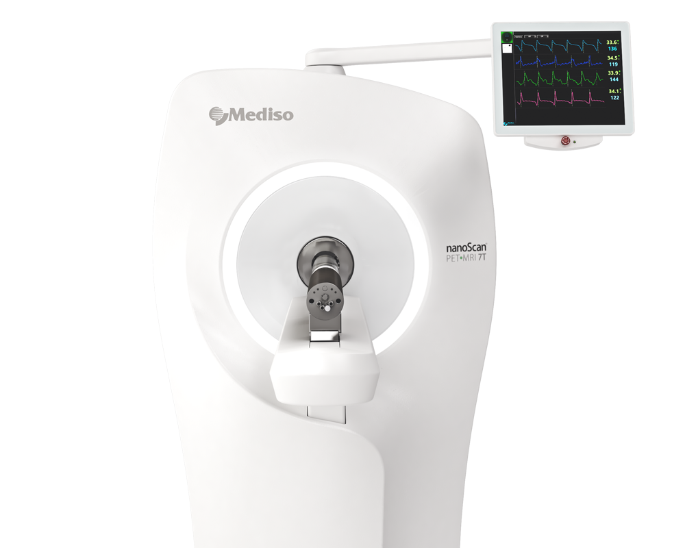
All the mice were scanned with a 3 T Mediso nanoScan® PET/MRI scanner (Mediso Ltd., Hungary) with a triple-mouse imaging chamber. Two 2-min anatomical T1 MR scans were performed prior to tracer injection (head receiver coil, matrix size 96 × 96 × 22, voxel size 0.24 × 0.24 × 0.80 mm3, repetition time 677 ms, echo time 28.56 ms, and flip angle 90°). The injected dose of [18F]PI-2620 delivered in 200 µl saline via intravenous injection was 12.7 ± 2.1 MBq. PET emission was recorded in a dynamic 0–60 min window. The frames used were 6 × 10, 2 × 30, 3 × 60, 5 × 120, 5 × 300, and 5 × 600. The list-mode data within the 400–600 keV energy window were reconstructed using a 3D iterative algorithm (Tera-Tomo 3D, Mediso Ltd., Hungary) with the following parameters: matrix size of 55 × 62 × 187 mm3, voxel size of 0.3 × 0.3 × 0.3 mm3, 8 iterations, and 6 subsets. Decay, random, and attenuation corrections were applied. The T1 image was used to create a body–air material map for attenuation correction. We longitudinally studied PS19 (n = 10) and age-matched wild-type mice (n = 10, WT; C57BL6) at 5.9, 7.7, 10.2, and 12.4 months of age. The sample size was selected based on the assumption of detecting a 10% difference between genotypes at the latest time point with a power of 0.8, applying an α of 0.05. No randomization was used to allocate the experimental units due to the absence of any intervention. No dropouts were registered; hence, all the mice were included in the subsequent analysis. Blinding was not applied during the scanning process, but it was implemented during image analysis, where an automatic coregistration step guaranteed reader independence. The normalization of the PET data was performed by calculating the volume of distribution (VT) images obtained from the full dynamic scan.
The MRI volumetric analysis was performed in a blinded manner on coronal sections by manual delineation of the cerebellum, the brainstem, and the striatum (each in three adjacent planes) using PMOD. We conducted a test–retest procedure to ensure the reliability of MRI segmentation, which displayed a high congruency (r) of > 0.9 across 10 test cases. The cerebellum and brainstem were considered as a combined hindbrain region.
- Regarding tau sensitivity [18F]PI-2620 tau PET imaging detected age-dependent increases in tau pathology in PS19 mouse models compared to wild-type mice. Strong correlations were observed between PET signals and AT8-positive tau pathology regions.
- The authors found that cellular sources of PET signals were primarily derived from neurons and oligodendrocytes with tau aggregation, while astrocytic tau did not significantly contribute to the signals. Autoradiography confirmed similar patterns in human samples.
- MRI volumetric changes showed that tau accumulation in the brainstem correlated with a decrease in hindbrain volume in PS19 mice (see Figure 3).

Figure 3 Cell sorting after radiotracer injection identifies neurons as the predominant origin of [18F]PI-2620 tau PET signals. e, f Quantitative correlation between brainstem tau PET signals and radioactivity per single neuron or astrocyte. g, h Data-driven voxelwise correlation between radioactivity per single neuron or astrocyte and [18F]PI-2620 tau PET images using statistical parametric mapping of the combined sample of PS19 and WT mice.
Conclusions
In this translational study, the authors used a large spectrum of methodological approaches, including innovative scRadiotracing and a cell-type-specific correlation of autoradiography signals, to disentangle the discrepant findings of previous reports that investigated second-generation tau PET in 4R-tauopathies. As a major achievement, they detected elevated radiotracer binding in isolated neurons after in vivo injection in mice. Furthermore, the data indicated that tau PET signals in individuals with 4R-tauopathies are driven by dense neuronal and oligodendroglial tau aggregation, whereas faint tau-positive structures of astrocytes and tau fragments are not capable of translating radiotracer binding into in vivo signals. The first [18F]PI-2620 PET-to-autopsy correlation was provided and showed that cortical tau PET signals deserve optimized target regions at the boundary between gray and white matter.
The overarching research question of this work addressed the validity of second-generation tau PET signals in individuals with 4R-tauopathies. In conclusion the presented novel approach of cell sorting after radiotracer injection can be readily used to test the cell type specificity of novel radiotracers with 4R-tau affinity.
Original link Springer Nature
How can we help you?
Don't hesitate to contact us for technical information or to find out more about our products and services.
Get in touch