Longitudinal positron emission tomography and postmortem analysis reveals widespread neuroinfammation in SARS-CoV-2 infected rhesus macaques
2023.07.29.
Juliana M. Nieuwland et al, Journal of Neuroinflammation, 2023
Summary
Background: Coronavirus disease 2019 (COVID-19) patients initially develop respiratory symptoms, but they may also sufer from neurological symptoms. People with long-lasting efects after acute infections with severe respiratory syndrome coronavirus 2 (SARS-CoV-2), i.e., post-COVID syndrome or long COVID, may experience a variety of neurological manifestations. Although we do not fully understand how SARS-CoV-2 afects the brain, neuroinfammation likely plays a role.
Methods: To investigate neuroinfammatory processes longitudinally after SARS-CoV-2 infection, four experimentally SARS-CoV-2 infected rhesus macaques were monitored for 7 weeks with 18-kDa translocator protein (TSPO) positron emission tomography (PET) using [18F]DPA714, together with computed tomography (CT). The baseline scan was compared to weekly PET–CTs obtained post- infection (pi). Brain tissue was collected following euthanasia (50 days pi) to correlate the PET signal with TSPO expression, and glial and endothelial cell markers. Expression of these markers was compared to brain tissue from uninfected animals of comparable age, allowing the examination
of the contribution of these cells to the neuroinfammatory response following SARS-CoV-2 infection.
Results: TSPO PET revealed an increased tracer uptake throughout the brain of all infected animals already from the frst scan obtained post-infection (day 2), which increased to approximately twofold until day 30 pi. Postmortem immunohistochemical analysis of the hippocampus and pons showed TSPO expression in cells expressing ionized calcium-binding adaptor molecule 1 (IBA1), glial fbrillary acidic protein (GFAP), and collagen IV. In the hippocampus of SARS-CoV-2 infected animals the TSPO+ area and number of TSPO+ cells were signifcantly increased compared to control animals. This increase was not cell type specifc, since both the number of IBA1+TSPO+ and GFAP+TSPO+
cells was increased, as well as the TSPO+ area within collagen IV+ blood vessels.
Conclusions: This study manifests [18F]DPA714 as a powerful radiotracer to visualize SARS-CoV-2 induced neuroinfammation. The increased uptake of [18F]DPA714 over time implies an active neuroinfammatory response following SARS-CoV-2 infection. This infammatory signal coincides with an increased number of TSPO expressing cells, including glial and endothelial cells, suggesting neuroinfammation and vascular dysregulation. These results demonstrate the long-term neuroinfammatory response following a mild SARS-CoV-2 infection, which potentially precedes long-lasting neurological symptoms.
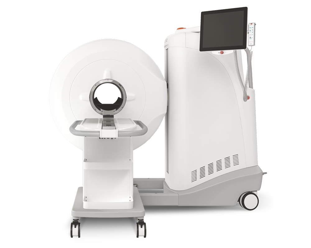
Results from MultiScan™ LFER150 PET/CT
Four Indian-origin male rhesus monkeys (Macaca mulatta) were infected with SARS-CoV-2, strain hCoV-19/Netherlands/NH-RIVM-27142/2021, the Delta variant via a combined intratracheal and intranasal route. The determination of the presence of SARS-CoV-2 mRNA and subgenomic mRNA in the nasal and throat swabs was performed using reverse transcription quantitative PCR.
A baseline PET–CT was acquired pre-infection (day-5) to set a reference parameter. Weekly PET–CTs were obtained using MultiScan LFER PET/CT The animals were positioned head-first supine. Following a scout-view, an intravenous bolus (1–2 ml) of approximately 180 MBq [18F]DPA714 was administered. All animals underwent the same scan procedure and number of scans, 8 scans total per animal. The PET image obtained 20–30 min post-injection was used for longitudinal analysis throughout the whole study. Afterwards a CT was acquired to use for attenuation correction. The system utilizes cone-beam CT technology which covers a volume of 150 × 200 × 200 mm3 in a single rotation of 32.4 s. For each scan a single rotation of 480 projections was captured. The main scan parameters applied for scans used in this manuscript were 80 kV, 720 μA and an exposure time of 0.09 s. The emission data were iteratively reconstructed (OSEM3D, 8 iterations and 9 subsets with an isotropic voxel size of 0.8 mm) into a single frame PET image normalized and corrected for attenuation, scatter, and random coincidences using the CT, and corrected for radioactive decay.
The analysis was performed with VivoQuant 4.5 (Invicro, Boston, USA). Based on the structures and regions available in the cortical hierarchy atlas of the rhesus monkey (CHARM) and the subcortical hierarchy atlas of the rhesus monkey (SARM) a selection of several regions of interest (ROIs) was made to fuse with the PET–CT data obtained within the study. standardized uptake values (SUV) of the ROIs were calculated resulting in an average signal of the ROI represented by the SUVmean and an average SUV within a 1-mm3 spherical volume around the voxels with the highest value by the SUVpeak.
- The PET/CT scans showed a continuous increase in tracer uptake over the course of infection throughout the brain (Fig. 2A).
- The average peak in increase was reached at day 30 pi, with an average SUVmean of 2.6, and afterwards the signal decreased (average SUVmean of 2.3 at day 44) but did not drop to the pre-infection level (SUVmean of 1.4 at day 0; Fig. 2B).
- The [18F]DPA714 uptake increased approximately twofold (range 1.4–2.0 fold) from day 2 to day 30 pi and demonstrated a significant correlation during the course of 44 days for all four animals (Fig. 2B)
- 44 days for all four animals (r = 0.905, p = 0.005) (Fig. 2B). When investigating the four infected animals separately, a similar increase in SUVmean was observed throughout the entire brain.
- SUVmean of ten ROIs of the left and right hemisphere were analyzed. No major differences were observed between regions of different hemispheres and all regions showed an increased signal, which was higher at day 30 or day 37 compared to day 44.
- Hippocampus and dorsal pons displayed a significant increase in tracer uptake (Fig. 2E) and correlation in SUVmean signal during the course of infection in all four animals
- The four animals individually also demonstrated an increase in SUVmean in these ROIs, with maximum SUVmean values of 2.6 (range 2.2–2.9) for the hippocampal formation (Fig. 2C) and 2.5 (range 2.2–2.7) for the dorsal pons (Fig. 2D).
- Three of the four animals (R1, R2 and R4) reached this maximum in SUVmean at day 30, however for both the hippocampal formation and dorsal pons R3 showed the highest SUVmean at the next scan obtained at day 37 (Fig. 2C, D).

Full article on jneuroinflammation.biomedcentral.com
How can we help you?
Don't hesitate to contact us for technical information or to find out more about our products and services.
Get in touch