Pretargeted PET Imaging with a TCO-Conjugated Anti-CD44v6 Chimeric mAb U36 and [89Zr]Zr-DFO-PEG5-Tz
2022.04.20.
Dave Lumen et al., 2022, Bioconjugate Chemistry
Summary
Quantitative positron emission tomography (PET) imaging can be used in preclinical as well as clinical research and provides important information about the pharmacokinetics of monoclonal antibodies (mAbs) and derivatives thereof, particularly with respect to the kinetics of tumor accumulation and washout from nontarget tissues. During the last decades, many antibodies have been developed for cancer diagnosis and treatment, and recent advances in the production of tailored antibodies for specific targets have provided several new radioimmunoconjugate candidates for immuno-PET imaging. These second-generation radioimmunoconjugates can be grouped into different categories: (i) antibody–drug conjugates (ADCs), designed to release a drug when reaching its target; (ii) multispecific mAbs, recognizing two or more targets; (iii) glycoengineered mAbs, which are modified to enhance the antibody-dependent cytotoxicity; and (iv) mAb fragments and nanobodies to tailor the radioimmunoconjugate pharmacokinetics. The relatively slow pharmacokinetics of antibodies require that the radioactive half-life of the isotope must be compatible with the biological half-life of the mAb. In practice, this means that for immuno-PET imaging the antibodies are often labeled with isotopes with long, even multiday physical half-lives such as 89Zr (78.41 h), 64Cu (12.70 h), and 124I (4.18 d), which allows for the detection of the radiolabeled antibodies after accumulation at the tumor and clearance from the circulation. It usually takes several days until nonbound antibodies are cleared from the circulation, and the optimal target-to-non-target (T:NT) values are obtained for imaging. The administered radioactive dose can therefore be high. The levels of radiolabeled mAbs in blood can be reduced using special clearing agents; however, this does not solve the problem of slow accumulation kinetics of mAbs in the tumor. Achieving high target-to-non-target values more rapidly would minimize the lag time needed between the radiotracer injection and the PET imaging, reducing exposure of the patient to radioactivity and the effective dose. Significant efforts have been dedicated to overcome these obstacles through the development of engineered antibody variants with faster pharmacokinetics and pretargeted approaches for radiolabeling the antibodies in vivo after their administration and peak accumulation to the target site. Recently, in vivo click reactions based on the bio-orthogonal inverse electron demand Diels–Alder ligation (IEDDA) between dienophile-functionalized antibodies and small-molecule radioligands based on tetrazine structures have obtained high interest. Pretargeted immuno-PET imaging would bring significant advantages: reducing the radioactive exposure of the patients and allowing the use of the short half-live radionuclides for imaging purposes (Figure 1). The preclinical proof of concept of the two-step pretargeted immuno-PET imaging and radioimmunotherapy with IEDDA have been successfully achieved by several research groups.
Bio-orthogonal click reactions are specific and selective reactions that can take place under physiological conditions and rapidly react even at low concentrations in vivo. Fast reaction kinetics and selectivity have made them a favorable choice for effective in vivo radiolabeling methods for pretargeted imaging and therapy. The IEDDA ligation between olefins or alkynes (e.g., trans-cyclooctene or TCO) and 1,2,4,5-tetrazines (e.g., tetrazine or Tz) is a selective, fast, high-yielding, biocompatible, and bio-orthogonal reaction, in which the reaction counterparts will undergo two concerted reactions to afford a coupling product under the formation of a pyridazine and dinitrogen (Figure 1). Reaction between TCO and Tz holds one of the fastest reaction kinetics from all click chemistry methods, which makes them ideal functional groups for in vivo applications. Rate constants for the reaction between tetrazine and TCO can exceed 100,000 M–1 s–1, orders of magnitude faster than either the Staudinger or strain-promoted azide–alkyne cycloaddition ligations. Rossin et al. used the IEDDA for the first time for pretargeted SPECT imaging, and the first pretargeted PET study was reported by Weissleder and Lewis. TCO isomerizes quickly to a less reactive cis-cyclooctene (CCO) in vivo unless conjugated to a macromolecule; therefore, most of the published pretargeting studies are based on the IEDDA ligation between a TCO-conjugated antibody and a small-molecular tetrazine carrying the radiolabel.
In this study, a 89Zr-labeled tetrazine ([89Zr]Zr-DFO-PEG5-Tz, [89Zr]Zr-3) was developed and utilized as a tool for investigation and comparison of targeted and pretargeted PET imaging of head-and-neck squamous cell carcinoma (VU-SCC-OE) xenografts using an anti-CD44v6 chimeric mAb (cmAb) U36. U36 was chosen for the study because it has shown high and selective tumor uptake in head-and-neck squamous cell carcinoma (HNSCC) patients and it internalizes into cells only to a limited extent. The splice variant v6 of the cell membrane glycoprotein CD44 (CD44v6) is expressed only in a few normal epithelial tissues (e.g., thyroid and prostate gland), but it plays a significant role in solid tumor growth and metastasis development. For the HNSCC, >96% of tumors show CD44v6 expression by at least 50% of the cells. In addition to squamous cell carcinomas, CD44v6 is overexpressed in adenocarcinomas and ovarian cancer and in hematological tumors. Expression of CD44v6 in tumors has been imaged by several research groups using U36 or its variants after radiolabeling it with different long-living radionuclides. In this study, U36 was conjugated with trans-cyclooctene and the conjugation ratio was optimized with biodistribution studies. TCO–U36 was radiolabeled in vitro and in vivo using [89Zr]Zr-3, and the uptake levels in VU-SCC-OE tumors were quantified with PET-CT/MRI and ex vivo biodistribution studies.
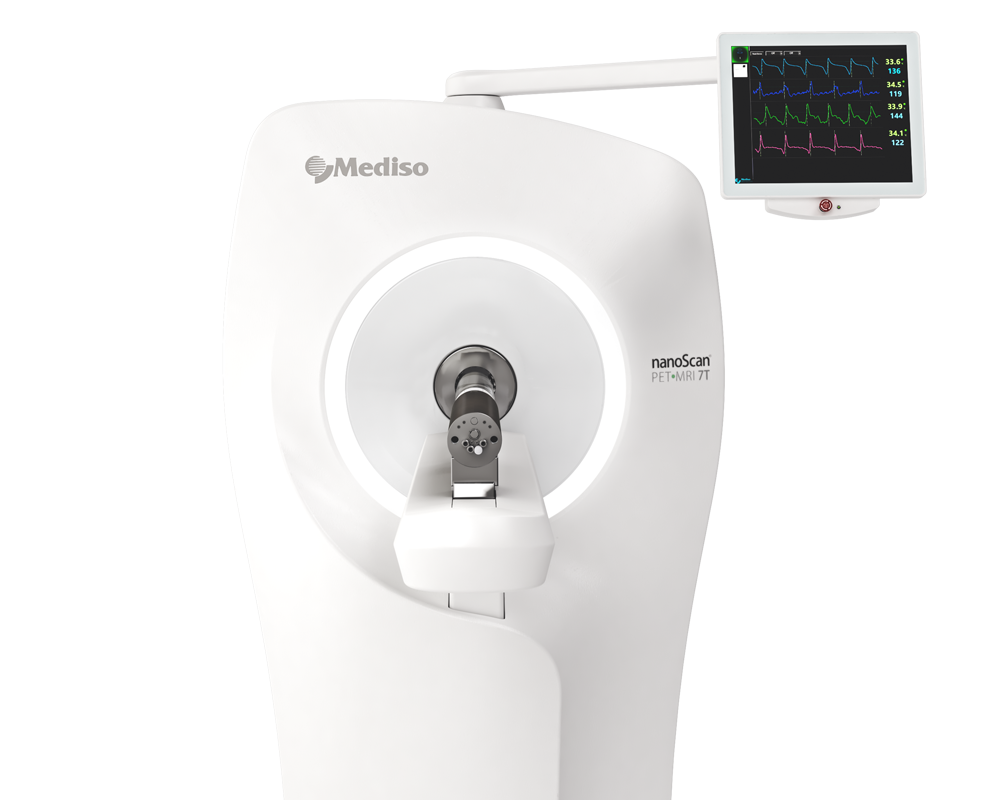
Results from the nanoScan PET/CT
For the biodistribution Study of In Vitro-Labeled U36–TCO and In Vivo Labeling of U36–TCO with [89Zr]Zr-3 (27:1 TCO-to-U36), Experiments were done in nude female mice (HSD:athymic nude Foxn1nu, 15–30 g; Charles River, Germany), aged 8–10 weeks at the time of the experiment, bearing subcutaneously implanted VU-SCC-OE xenografts (tumor volumes varied from 205 to 914 mm3). Mice were randomized to the three groups (n = 4/group): group 1 received the in vitro-labeled [89Zr]Zr-3–TCO–U36 and groups 2 and 3 for the pretargeted approach received [89Zr]Zr-3 24 and 48 h after U36–TCO administration. On day 1, group 1 mice were injected (i.v.) with in vitro-labeled [89Zr]Zr-3–TCO–U36 (4.4 ± 0.4 MBq, 0.1 mg, 0.66 nmol) and groups 2 and 3 mice were injected (i.v.) only with U36–TCO (0.1 mg, 0.66 nmol). For group 2, [89Zr]Zr-3 (4.1 ± 0.3 MBq, 0.7 μmol, 0.66 nmol) was injected (i.v.) 24 h after U36–TCO injection and for group 3 (3.9 ± 0.5 MBq, 0.7 μmol, 0.66 nmol) (i.v.) 48 h after U36–TCO injection. Group 1 mice were imaged with PET-CT/MRI at 1 (dynamic scan), 24, 48, and 71 h after U36 injection, group 2 mice were imaged 1 (dynamic scan), 24, and 47 h, and group 3 were imaged 1 (dynamic scan) and 23 h after the injection of the tracer. All mice were sacrificed at 72 h p.i. of the U36 injection, and the collected organs (urine, blood, gall bladder, pancreas, spleen, kidney, liver, heart, lung, stomach, small intestine, large intestine + cecum, feces (1–2 pellets from the rectum), bladder, skeletal muscle, bone (tibia), bone (skull), brain, skin, and head) were weighted and the amount of radioactivity in each tissue was measured by a γ-counter. Radioactivity uptake was calculated as the percentage of the injected dose per gram of tissue (% ID/g). Quantitative PET image analysis was performed by defining regions of interest (ROIs) around the tumor with CT or MRI as the anatomical reference. Radioactivity concentration was expressed as an SUV, calculated using the average radioactivity concentration of the ROI normalized with the injected radioactivity dose and animal weight.
For the biodistribution Study of In Vitro-Labeled TCO–U36 and In Vivo Click Reaction (6:1 TCO-to-U36).
Figure 7. shows coronal PET/CT images for all groups at 71 h p.i. of the U36 antibody administration in VU-SCC-OE xenografts. Despite the lower activity concentration in the pretargeted tumors, the tumors were clearly visible by PET-computed tomography (PET/CT) due to the low background activity (Figure 7). Tumor activities were quantified by delineating region of interests around the tumors and by calculating standardized uptake values (SUVs) for all groups at 1, 24, 48, and 71 h after the U36 injection (Figure 8). The tumor volumes varied from 31 to 793 mm3, and the heterogeneous structure of the tumors caused some additional challenge for the image analysis and calculation of the SUVs. Due to the structural heterogeneity (necrotic core poorly perfused), the activity concentrations varied significantly between the tumors, resulting in high variation of the SUVs between tumors from the same group. In general, small tumors (<100 mm3) had clearly higher activity concentration compared to the larger ones.
- Anti-CD44v6 monoclonal antibody U36 was successfully used for 89Zr-immuno-PET imaging of head-and-neck squamous cell carcinoma xenograft tumors using both a targeted and pretargeted approach.
- The authors' results demonstrate that the pretargeting of TCO–U36 with the tetrazine [89Zr]Zr-3 constitutes a promising concept for in vivo pretargeted PET imaging on antibodies with zirconium-89 and warrants further investigation into radiolabeling of 3 with shorter-lived PET radionuclides like 68Ga.
- An alternative and potential method for in vitro radiolabeling of 89Zr-labeled radioimmunoconjugates is presented using IEDDA and [89Zr]Zr-3.
How can we help you?
Don't hesitate to contact us for technical information or to find out more about our products and services.
Get in touch
