Radiation dosimetry of Tc-99m-PSMA I&S: a single-center prospective study
2020.12.04.
Szabolcs Urbán et al., Journal of Nuclear Medicine, 2020
Abstract
Introduction: Tc-99m-labeled Mas3-y-nal-k(Sub-KuE) (Tc-99m-PSMA I&S) is a prostate specific membrane antigen (PSMA) tracer that can be used for planar and SPECT/CT gamma imaging and radioguided surgery (RGS). The primary aim of this study was to estimate the dosimetry of Tc-99m-PSMA I&S using a hybrid method (sequential gamma planar imaging and one single SPECT/CT) in healthy volunteers. The secondary aim was to depict the tracer biodistribution and tumor-to-background ratios (TBR) in patients with prostate cancer (PCa).
Methods: Dosimetry of Tc-99m-PSMA I&S was investigated in four healthy volunteers. Whole-body planar imaging was acquired at 1, 2, 3, 6 and 24 hours, and SPECT/CT at 6h after tracer injection. Contours of organs were drawn on all acquisitions to determine organ activity at each timepoint. Absorbed dose was estimated using two methods: 1) independent curve-fitting manual method (Levenberg-Marquardt-based algorithm using dose factors from Radiation Dose Assessment Resource (RADAR) web site) and 2) OLINDA/EXM® v 2.0 software (HERMES Medical Solutions). Biodistribution of Tc-99m-PSMA I&S was assessed in ten patients with PCa on SPECT/CT images at 6 h. Tumor uptake (SUVmax), and TBR (tumor SUVmax/ background organ SUVmean) using muscle (T/M), bladder (T/B) and intestine (T/I) as background organs were determined.
Results: The mean injected activity of Tc-99m-PSMA I&S was 717 MBq (range: 562-828). No adverse events related to the injection of Tc-99m-PSMA-I&S were reported. The average radiation effective dose was 0.0055 mSv/MBq with the RADAR manual method and 0.0052 mSv/MBq with OLINDA/EXM®. Total body effective dose ranged between 3.33-4.42 and 3.11-4.23 mSv, respectively. All PCa patients showed high tracer uptake in primary and metastatic lesions with T/M, T/B, T/I ranging from 5.29 - 110, 0.11 – 7.02 and 0.96 – 16.30, respectively.
Conclusion: Effective doses of Tc-99m-PSMA I&S were comparable to those known for most of the Tc-99m tracers and was lower than 68Ga-labelled and 18F-labelled agents. Tc-99m-PSMA I&S SPECT/CT showed high TBR in PCa patients. This study can provide required data for translation and approval of Tc-99m-PSMA I&S by regulatory agencies.

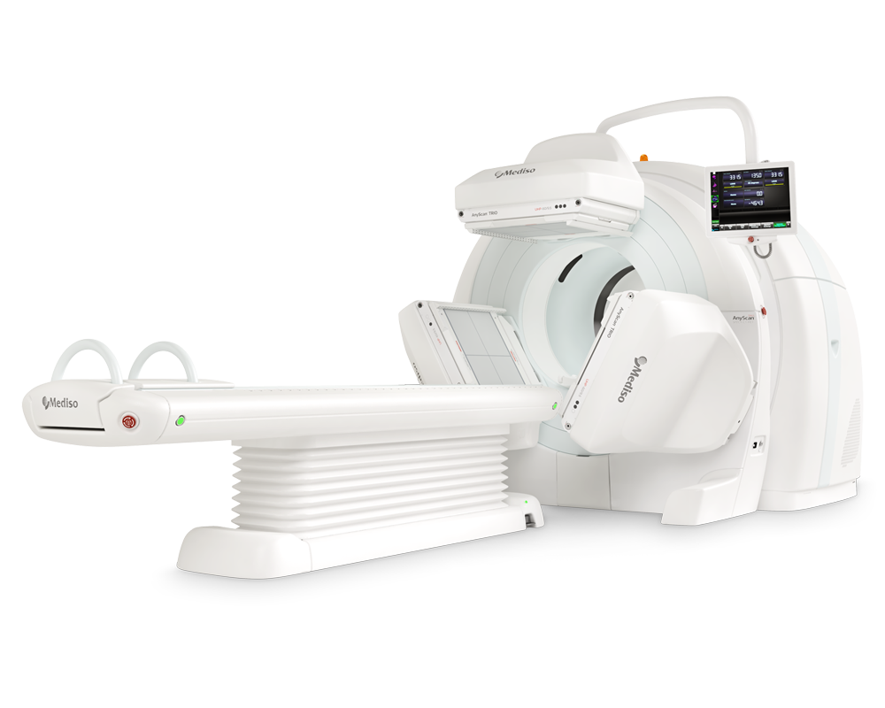
Acquisition with AnyScan® TRIO SPECT/CT
A hybrid imaging method was used to determine the dosimetry of Tc-99m PSMA I&S: multiple time-point whole-body (WB) planar imaging and one single quantitative SPECT/CT scan. In all healthy subjects (n=4) WB anterior and posterior scintigraphy was performed at 1, 2, 3, 6 and 24 h after radiopharmaceutical administration (Figure 1)

using a triple head gamma camera (AnyScan Trio SPECT/CT, Mediso Medical Imaging Systems Ltd.), equipped with low energy, high-resolution collimators (LEHR). The scanning speed was 18 cm/min, matrix size of 256 × 1024 pixels was used and a symmetric 20% window was set at 140 keV. In all volunteers and PCa patients (n=14) whole-body (mid-thighs to vertex) SPECT/CT images were acquired at 6 h after radiopharmaceutical administration (Figure 2).

Quantitative SPECT images were acquired using a 128 × 128 matrix with a 20% energy window centered at 140 keV with adjacent scatter correction windows. A total of 96 projection views were acquired over 360 degrees in 3.75 degree steps at 10 s per view. The number of bed positions was 3D Reconstruction of raw SPECT data was carried out using the iterative Tera-Tomo 3DTM (Mediso Medical Imaging Systems Ltd.) software which is based on the order of sets and subsets (48 iterations/4 subsets) method. CT-based attenuation correction and Point Spread Function (PSF) correction were used. CT images were acquired using low dose CT (120 KeV, 100 mAs, 1.5 pitch factor and 2.5 mm slice thickness). To improve the visibility of the gastrointestinal tract, Macrogol 1500 (50g/l) was administered orally one hour before SPECT/CT imaging.
Results from AnyScan® TRIO SPECT/CT
The median injected activity of Tc-99m PSMA I&S was 695 MBq (range 553-821). No adverse events related to the injection of Tc-99m-PSMA-I&S were reported. High uptake of Tc-99m-PSMA I&S was observed 6 hours after injection both in primary tumors (10/10 patients (100%), mean SUVmax 13.37 (range 3.25–44.00), and in metastatic lesions (3/10 patients (30%), mean SUVmax 5.71 (range 1.80–8.48) (Table 3).

The mean T/M, T/B, T/I ratio in the primary tumors was 30.22 (range 7.95-110.00), 1.59 (range 0.11-7.02), 5.56 (range 0.96-16.30) and in the metastasis 14.97 (range 5.29-24.23) 0.60 (range 0.33-0.92), 3.16 (range 1.20-4.82), respectively. Figure 4 shows pathological tracer uptake in primary PCa (#012), and in patients with metastases in bone (#011) and lymph nodes (#014). Based on the SPECT counts, the average activity of the pathological lesions 6 hours after tracer injection was 0.16 MBq (range 0.01-0.96). Based on the physical half-life of Tc-99m, the average lesion activity after 24 and 48 hours was estimated to be 19.73 kBq (range 1.25-120.00) and 1.23 kBq (range 0.08-7.55), respectively.
Full article on jnm.snmjournals
-
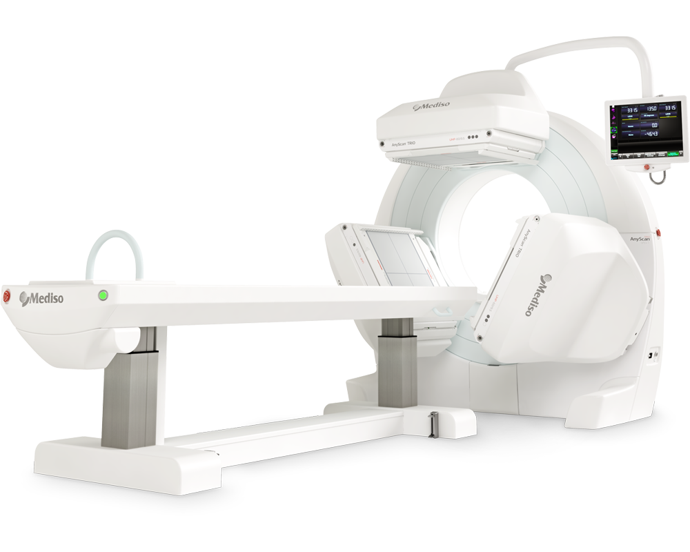
AnyScan® TRIO SPECT
Ultra-fast Triple-NaI-Detector SPECT for wide range of clinical applications
-
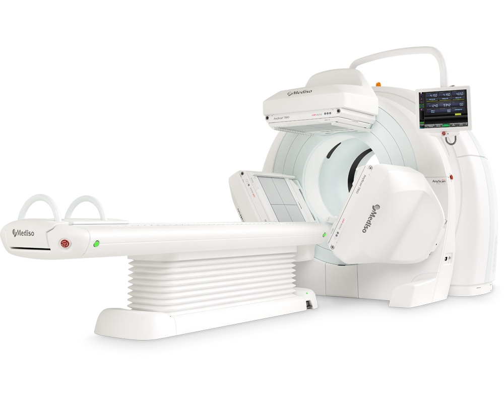
AnyScan® TRIO SPECT/CT
Ultra-fast Triple-NaI-Detector SPECT system for wide range of clinical applicati...
-
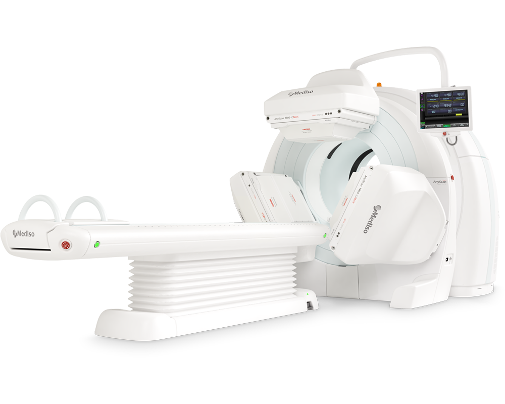
AnyScan® TRIO SPECT/CT IQMAX
Diagnostic and Theranostic Image Quality at MAXimum Level
-
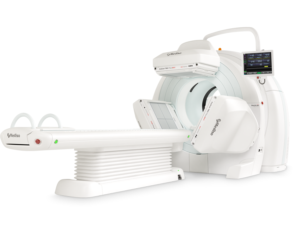
AnyScan® TRIO SPECT/CT TheraMAX
Theranostic and Diagnostic Imaging with MAXimum Performance
-
AnyScan® TRIO SPECT/CT/PET
An integrated triple-detector SPECT/CT and PET/CT system in a single-room instal...
-
InterView™ XP Processing Software
Planar, Whole-Body & SPECT Processing Software


How can we help you?
Don't hesitate to contact us for technical information or to find out more about our products and services.
Get in touch