Radiolabeled F(ab′)2-cetuximab for theranostic purposes in colorectal and skin tumor-bearing mice models
2018.05.17.
P.-S. Bellaye et al., Clinical and Translational Oncology, 2018
Summary
The epidermal growth factor receptor (EGFR) has evolved over the years into a main molecular target for the treatment of different cancer entities. EGFR is a glycosylated transmembrane protein involved in regulating cell growth, differentiation and survival of malignant cells. This receptor is often overexpressed in various malignancies such as head and neck squamous cell carcinoma (HNSCC), gastrointestinal/abdominal carcinoma, lung and reproductive tract carcinomas, melanomas, glioblastomas and thyroid carcinoma. Despite some controversies, the overexpression of EGFR is often associated with a poor clinical prognosis and resistance to radiation therapy. Therefore, therapeutic strategies involving monoclonal antibodies against EGFR, such as cetuximab, have been used pre-clinically and clinically. Cetuximab is a chimeric monoclonal antibody, which binds specifically with a high affinity to the extracellular domain of the EGFR. Cetuximab is used alone or in combination and its therapeutic indications are (i) colorectal and HNSCC and (ii) HNSCC with external radiotherapy. While the combination of cetuximab with radiotherapy showed improved survival tumor response remains heterogeneous and cetuximab failed to show benefit over chemoradiotherapy. Hence, radioimmunotherapy based on cetuximab labeled with therapeutic radionuclides appears as a promising strategy allowing the delivery of radiation dose specifically to tumor cells expressing high level of EGFR while sparing normal tissues.
In this regard, theranostic strategy based on monoclonal antibodies and their deriving structures could represent an exciting approach. It is possible to convert a purely imaging probe into a single-entity theranostic agent by simply switching the radionuclide from a γ-emitter (e.g.: 111In) to a β-emitter (e.g.: 90Y) or using a radionuclide gathering both imaging and therapeutic properties (e.g.: 177Lu). The relevance of using cetuximab radiolabeled with 111In in diagnostic imaging has been demonstrated in several animal models mainly due to its high tumor targeting property and its adequate half-life. 177Lu is a theranostic radionuclide of choice to target small tumors or metastatic deposits due to the medium energy of the emitted beta particles and their tissue penetration of 1.5 mm. In addition, 177Lu emits γ-rays which allow diagnostic imaging.
A major concern with the use of a β-emitter such as 177Lu as radionuclide is the selection of a chelating agent that forms a sufficiently stable complex to prevent in vivo loss of the radiometal. To circumvent the drawbacks of chelating agents, it is possible to generate fragments [Fab and F(ab′)2] of the monoclonal antibody to improve its penetration within the tumor. These fragments can be prepared through enzymatic cleavages [papain for Fab and pepsin for F(ab′)2] or by genetic engineering. Given their pharmacokinetic properties, F(ab′)2 (110 kDa) could be valuable theranostic agents.
In the present work, the authors' aims were to design F(ab′)2-cetuximab-based theranostic agents with both diagnostic and therapeutic capabilities and to assess them in murine preclinical cancer models. First, they characterized in vitro the F(ab′)2 fragment of cetuximab radiolabeled with 111Indium (111In-DOTAGA-F(ab′)2-cetuximab) in comparison with whole cetuximab used as a reference (111In-DOTAGA-cetuximab). Then, evaluated the stability of DTPA- and DOTAGA-radiolabeled F(ab′)2-cetuximab.
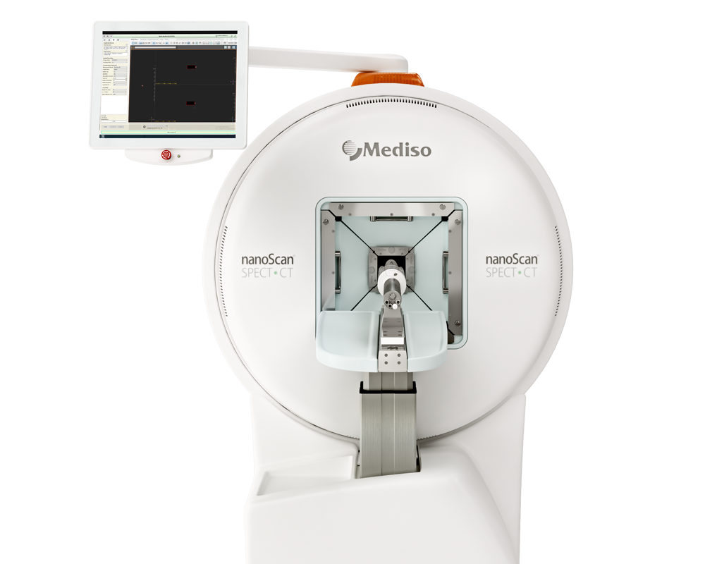
Results from nanoScan SPECT/CT
For the imaging studies, female Balb/c nu/nu mice (n = 3, 6–8 weeks old, purchased from Charles River, France) were grafted by subcutaneous injection of colon tumor fragments from human patients (CR-LRB-014P). When grown these tumors were collected and fragments from these tumors were implanted into a second set of Balb/c nu/nu mice (6–8 weeks old). 3 or 5 weeks after tumor implantation, tumor-bearing mice were given 25 μg 111In-DOTAGA-F(ab′)2-cetuximab (13–15 MBq) by intravenous injection. In a second experiment to assess the specificity of the targeting in vivo, a group of mice received 25 μg 111In-DOTAGA-F(ab′)2-cetuximab (3–3.5 MBq) in co-injection with excess (2500 μg) cold-F(ab′)2-cetuximab. Two groups of mice were then studied: (1) 111In-DOTAGA-F(ab′)2-cetuximab and (2) 111In-DOTAGA-F(ab′)2-cetuximab + non radiolabelled-DOTAGA-F(ab′)2-cetuximab. SPECT/CT dual imaging was performed 3, 6, 20, 24, 48, and 72 h after the injection of the radiolabeled conjugate using the NanoSPECT/CT small animal imaging tomographic γ-camera. Mice were anaesthetized with isoflurane (1.5–3% in air) and positioned in a dedicated cradle. CT and SPECT acquisitions were performed in immediate sequence. CT acquisitions (55 kVp, 34 mAs) were first acquired during 15–20 min, followed by helical SPECT acquisitions with 90–120 s per projection frame resulting in acquisition times of 45–60 min. Both indium-111 photopeaks (171 and 245 keV) were used with 10% wide energy windows.
Figure 2. shows the in vivo tumor specificity of 111In- DOTAGA-F(ab′)2-cetuximab. 111In-DOTAGA-F(ab′)2-cetuximab biodistribution was evaluated in Balb/c nude mice grafted subcutaneously with CR-LRB-014P human colon tumor fragments at 3, 6, 20, 24, 48 and 72 h post injection. Liver and kidney were the normal tissues with highest uptake (Fig. 2a). The liver uptake remained stable from 3 to 24 h post injection. Then, it started to significantly decrease at 48 and 72 h post injection. The kidney uptake significantly increased at 20 and 24 h compared to 3 h post injection and remained stable up to 72 h post injection. The bladder uptake remained stable throughout the experiment (Fig. 2a). Interestingly, the tumor uptake started to significantly increase at 20 h post injection and remained increased up to 72 h compared to 3 h post injection (Fig. 2a, b). Mice that were pre-injected with an excess of unlabeled F(ab′)2-cetuximab had significantly lower tumor uptake demonstrating the specificity of F(ab′)2-cetuximab for the CR-LRB-014P tumors (Fig. 2c).

Figure 3. shows that the 111In-DOTAGA-F(ab′)2-cetuximab is a reliable tool to monitor 17-DMAG treatment efficacy. Balb/c nude mice grafted subcutaneously with human primary colon tumor fragments (CR-LRB-014P) received 17-DMAG three times a week from D30 up to D58. Weight loss and tumor growth were monitored throughout the experiment. Mice received 111In-F(ab′)2-cetuximab every week from D30 (D38, D44, D51 and D58) for SPECT-CT imaging (imaging 24 h after injection). 17-DMAG induced a slight weight loss from D42 (12 days after treatment start) that was partially recovered by D58 compared to mice receiving vehicle (Fig. 3a). Interestingly, 17-DMAG induced a significant decrease in tumor growth compared to vehicle from D44 up to D58 (Fig. 3b). In parallel, tumor uptake of 111In-F(ab′)2-cetuximab measured by SPECT-CT imaging demonstrated that 17-DMAG treatment reduced tumor uptake compared to vehicle. Although only significant from D58, a marked reduced tumor uptake was observed from D44 (p = 0.059, Fig. 3c). Interestingly, tumor volume measured by SPECT-CT imaging reflected the decrease in tumor growth induced by 17-DMAG observed by manual measurements (Fig. 3d–f). However, a significant difference in tumor volume measured by SPECT-CT between 17-DMAG and vehicle was only observed at D51 while already observed at D44 by manual measurements (Fig. 3d). However, the decrease in tumor volume measured by SPECT-CT was clearly observed from D44 even though not significant (p = 0.065). Interestingly, there was a strong positive correlation between tumor volumes measured manually and with SPECT/CT imaging (Fig. 3e).

Figure 4. shows that 177Lu-DOTAGA-F(ab′)2-cetuximab prevents tumor growth in vivo. SWISS nude mice grafted subcutaneously with A431 cells received a single injection of 177Lu- DOTAGA-F(ab′)2-cetuximab (iv, 2, 4 or 8 MBq) on D14 after tumor inoculation. Control mice received vehicle on D14 (iv). While both 2 MBq and 8 MBq of 177Lu-DOTAGA-F(ab′)2-cetuximab did not induce weight loss in treated animals, 4 MBq induced a rapid weight loss at D20 which was normalized by D23 (Fig. 4a). Interestingly, all three doses of 177Lu-DOTAGA-F(ab′)2-cetuximab induced a reduction of tumor growth between D14 and D20 although only significant in mice receiving 4 and 8 MBq of 177Lu-DOTAGA-F(ab′)2-cetuximab (Fig. 4b, c). Taken together these results demonstrate that 177Lu-DOTAGA-F(ab′)2-cetuximab is able to prevent A431 cells’ tumor growth with a highest efficacy at 4 and 8 MBq.

- The authors have demonstrated that DOTAGA-F(ab′)2-cetuximab exhibits high in vivo and in vitro stability after radiolabeling with 111In and 177Lu.
- 111In-DOTAGA-F(ab′)2-cetuximab is a reliable tool for SPECT imaging of colorectal cancer overexpressing HER1 and enables accurate therapy efficacy monitoring (e.g. anti-HSP90 therapy).
- The radionuclide switch from 111In to 177Lu to form 177Lu-DOTAGA-F(ab′)2-cetuximab allows radioimmunotherapy coupling therapeutic efficacy and accurate imaging properties to treat and image HER1 expressing colorectal tumors.
How can we help you?
Don't hesitate to contact us for technical information or to find out more about our products and services.
Get in touch
