Imaging of fibrogenesis in the liver by [18F]TZ-Z09591, an Affibody molecule targeting platelet derived growth factor receptor β
2023.09.21.
Olivia Wegrzyniak et al., 2023, EJNMMI Radiopharmacy and Chemistry
Summary
Fibrogenesis is a dynamic process characterized by the ongoing accumulation of extracellular matrix (ECM), which can lead to the dysfunction or failure of the affected organ. Fibrotic diseases can occur in any organ, including the liver. Liver fibrosis can have several etiologies, but today those of metabolic origin are considered an important societal challenge because of the increasing prevalence of obesity and diabetes.
Today, liver fibrosis is rarely diagnosed before an advanced stage due to the lack of significant clinical signs associated with a moderate stage. Unfortunately, no anti-fibrotic therapies are currently approved in the case of severe liver fibrosis. Recently, efforts have been made to develop non-invasive tests, such as serum assay (e.g. FibroTest), imaging tests with, for instance, elastography devices (e.g. FibroScan) or multifactorial scoring systems (e.g. AST/platelet ratio index (APRI) and the Fibrosis Index Based on 4 Factors (FIB-4)). Unfortunately, these methods are still limited by their low sensitivity and specificity, especially at an early stage of the disease. The gold standard for assessing fibrosis is liver biopsy and its unavoidable limitations (invasiveness, complications, sampling variability, etc.). Therefore, the development of a non-invasive technique to quantify fibrogenesis would potentially be a major step forward in providing a more reactive and sensitive assessment of the activity of antifibrotic therapies.
Hepatic stellate cells (HSCs) when activated (aHSCs) play a major role in liver fibrogenesis as they are known to be the main source of ECM in liver fibrosis. There is an interest in imaging aHSCs as such an approach has the potential for diagnosis at an early stage, before pathological changes, prognosis, and monitoring reactively the response to treatment.
Platelet-derived growth factor receptor beta (PDGFRβ) is a biomarker of aHSCs but is absent on resting HSCs, it is thus a biomarker of fibrogenesis in the liver. The Affibody molecule Z09591 targeting PDGFRβ with a subnanomolar affinity was previously developed. Affibody molecules are small molecules of 58-amino-acid (6.5 kDa) three-helical bundle polypeptide. They are robust, easy to engineer, and small relative to e.g. antibodies, which is favorable for diagnostic imaging due to their rapid biodistribution and tissue penetration. The Affibody molecule Z09591 was previously labelled with a dodecane tetraacetic acid (DOTA) chelator and evaluated for the molecular imaging of PDGFRβ-expressing xenografts with [111In]DOTA-Z09591 and [68 Ga]DOTA-Z09591. However, labeling with fluorine-18 would be of interest since it has ideal nuclear properties for positron emission tomography (PET) imaging, allowing for e.g. high-resolution images as well as facile logistics due to its two-hour radioactive half-life.
Hence, given the implication of PDGFRβ in fibrogenesis and given the characteristics of the Affibody molecule Z09591, the authors speculated that the radiolabeled Affibody molecule targeting PDGFRβ could be suitable for detecting liver fibrogenesis. To further this hypothesis, the Affibody molecule Z09591 was conjugated to a trans-cyclooctene (TCO) group and labeled to a Flourine-18 labeled tetrazine (TZ) group, the resulting radiotracer [18F]TZ-Z09591 was evaluated in vitro on liver biopsies from patients with liver fibrosis and in vivo using the carbon tetrachloride (CCl4) mouse model of liver fibrosis.
Results from the nanoScan PET/CT
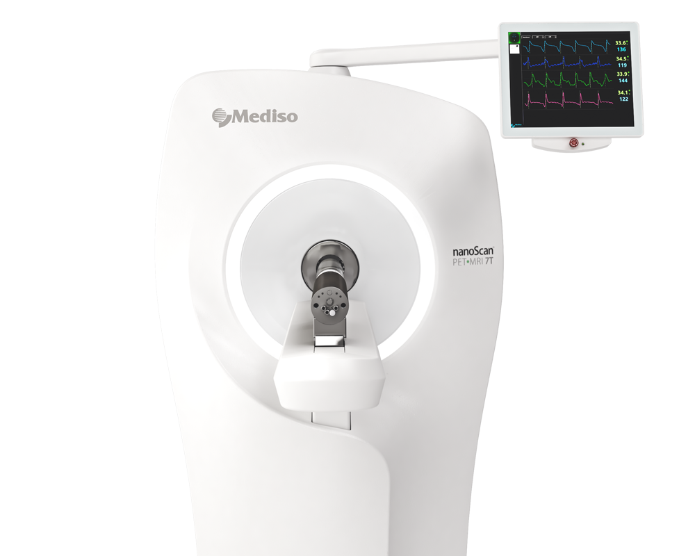
For the imaging studies, each animal was anesthetized (0,6L/min of 3% sevoflurane in 50/50% oxygen/medical air,) and placed in prone position on the bed of a preclinical PET/MRI scanner (nanoScan PET/MRI, 3T field strength, 10 cm field of view, Mediso). A heated bed supported body temperature, maintaining it at 37°C. Positioning was assisted by a scout MRI scan (GRE MultiFOV scout). [18F]TZ-Z09591 (10.4 ± 1.3 MBq) was administered as a bolus through an intravenous catheter in the tail. At the same time, scanner acquisition was started and continued for 150 min. The PET protocol was designed to provide repeated whole-body passes of increasing duration (3 beds per pass; 2 × 5 min, 2 × 10min, 4 × 30 min). After the scan, the animal was euthanized and whole-body MRI images were acquired for anatomical co-registration (SE MultiFOV).
Whole-body PET/MRI images were visualized and analyzed using the PMOD software (PMOD Technologies). Briefly, tissues of interest (brain, blood, lung, liver, spleen, kidneys, urine bladder, bone, bone marrow, muscle, small intestine, lower and upper large intestine (lumen/ content was included for each intestinal segment), stomach content) were segmented on summarized PET images and MRI projections, and the PET uptake value for each time point read out as SUVmean. The entire body was also segmented.
The ex vivo biodistribution (Fig. 4C) in healthy rats showed that [18F]TZ-Z09591 exhibited fast blood clearance with SUV values below one after 20 min. Kidney and urine uptakes were elevated at all time points. This was also observed in PET images (Fig. 4A, B). Liver uptake was high at an early time with a SUV of 4,07 at 5 min (Fig. 4C). However, the liver uptake decreased with time, at 20min the observed SUV was 0,64 ± 0,34, and 0,20 ± 0,10 at 240 min. This observation was the same as that obtained using dynamic PET (n = 4) (Fig. 4A, B). In vivo biodistribution based on dynamic PET shows that the liver revealed a decreasing uptake, from an SUV of 3,37 ± 0,39 at 5 min to 0,14 ± 0,05 at 152 min.
- The authors' data describe the evaluation of the PDGFRβ binding Affibody molecule Z09591 in the context of liver fibrosis. This strongly suggests that the PET tracer [18F]TZ-Z09591 specifically visualizes PDGFRβ expression, and thus aHSCs responsible for fibrogenesis, in a preclinical liver fibrosis model as well as in human tissue.
- Therefore, [18F]TZ-Z09591 appears to be a suitable candidate for PET imaging of liver fibrogenesis.
- Further prospective studies are required to evaluate the clinical utility of PET imaging of PDGFRβ expression using the [18F]TZ-Z09591 for the clinical assessment of liver fibrosis progression or regression based on these promising preliminary results.
Hogyan segíthetünk Önnek?
További termékinformációkért, vagy támogatásért keresse szakértőinket!
Vegye fel a kapcsolatot