Experimental Therapy of HER2-Expressing Xenografts Using the Second-Generation HER2-Targeting Affibody Molecule 188Re-ZHER2:41071
2022.05.20.
Yongsheng Liu et al, Pharmaceutics, 2022
Summary
HER2-targeted radionuclide therapy might be helpful for the treatment of breast, gastric, and ovarian cancers which have developed resistance to antibody and antibody-drug conjugate-based therapies despite preserved high HER2-expression. Affibody molecules are small targeting proteins based on a non-immunoglobulin scaffold. The goal of this study was to test in an animal model a hypothesis that the second-generation HER2-targeting Affibody molecule 188Re-ZHER2:41071 might be useful for treatment of HER2-expressing malignant tumors. ZHER2:41071 was efficiently labeled with a beta-emitting radionuclide rhenium-188 (188Re). 188Re-ZHER2:41071 demonstrated preserved specificity and high affinity (KD = 5 ± 3 pM) of binding to HER2-expressing cells. In vivo studies demonstrated rapid washout of 188Re from kidneys. The uptake in HER2-expressing SKOV-3 xenografts was HER2-specific and significantly exceeded the renal uptake 4 h after injection and later. The median survival of mice, which were treated by three injections of 16 MBq 188Re-ZHER2:41071 was 68 days, which was significantly longer (<0.0001 in the log-rank Mantel-Cox test) than survival of mice in the control groups treated with vehicle (29 days) or unlabeled ZHER2:41071 (27.5 days). In conclusion, the experimental radionuclide therapy using 188Re-ZHER2:41071 enabled enhancement of survival of mice with human tumors without toxicity to the kidneys, which is the critical organ.
Results from nanoScan® SPECT/CT
Biodistribution of 188Re-ZHER2:41071 in tumor bearing mice was measured in female BALB/C nu/nu mice. To establish the HER2-positive and HER2-negative xenografts, approximately 107 SKOV-3 cells or Ramos cells, respectively, were subcutaneously implanted in the hind legs of mice.
The average SKOV-3 tumor weight was 0.16 ± 0.12 g. The average Ramos tumor weight was 1.19 ± 0.62 g. Drinking water was supplemented with 5% sodium iodide 24 h before injection of 188Re-ZHER2:41071. Mice were injected intravenously with 10 µg (270 kBq) 188Re-ZHER2:41071 per mouse in 100 µL 0.9% sterile saline. At 1, 4, 8, 24, and 48 h after injection, one group of mice with SKOV-3 was euthanized and the biodistribution was measured. To test in vivo specificity, biodistribution and tumor uptake was measured 4 h after injection in one group of mice with Ramos xenografts. In vivo imaging was performed to confirm the biodistribution data.
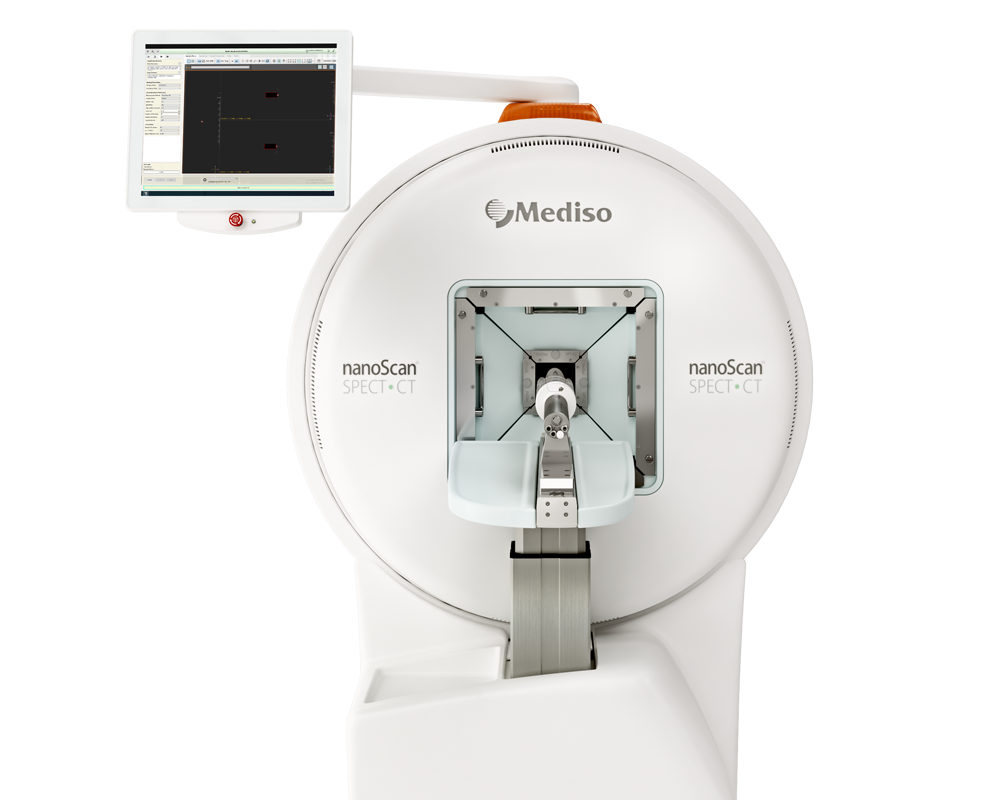
Two mice with SKOV-3 xenografts and one mouse with Ramos xenografts were injected with 4 MBq (10 µg) of 188Re-ZHER2:41071. The imaging was performed 1 h and 4 h p.i. for mice bearing SKOV-3 xenografts and 4 h for mice bearing Ramos xenografts using nanoScan SPECT/CT (Mediso Medical Imaging Systems, Budapest, Hungary). The data were reconstructed using Tera-Tomo™ 3D SPECT Software.
To evaluate the therapeutic efficacy of 188Re-ZHER2:41071, thirty female BALB/C nu/nu mice were subcutaneously implanted on the abdomen with 107 SKOV-3 cells. One week after the implantation, the mice were randomly distributed to three groups, A–C (10 mice per group). In group A (treatment with radiolabeled Affibody® molecule), mice were injected with 5 µg (16 MBq) of 188Re-ZHER2:41071 in 150 µL of 10% ethanol in 0.9% saline. In group B (treatment with non-labeled Affibody molecule), mice were injected with 5 µg of ZHER2:41071 in 150 µL of 10% ethanol in 0.9% saline. In group C (vehicle-treated control), mice were injected with 150 µL of 10% ethanol in 0.9% saline. Three injections separated by 48 h were performed intravenously. The tumor volumes at the start of the treatment (day 0) were 101 ± 28, 85 ± 42, and 86 ± 37 mm3 for mice in group A, B, and C, respectively. Throughout the experiment, tumor volumes and body weights were measured twice per week. For imaging HER2 expression during experimental therapy, SPECT/CT scans of mice bearing SKOV3 xenografts were performed using nanoScan SPECT/CT (Mediso Medical Imaging Systems, Budapest, Hungary). Two mice from each group were injected with 99mTc-labeled ZHER2:41071 Affibody® molecule (5 µg, 16 MBq) and imaging was performed at 4 h p.i.
- 188Re-ZHER2:41071 cleared rapidly from blood and normal tissues including kidneys. The uptake in tumor was high, 31 ± 4%ID/g already 1 h after injection. The tumor uptake remained to be high over time while the activity in kidneys declined rapidly. Already 4 h after injection, the tumor uptake was more than 8-fold higher than the renal uptake. This created a favorable precondition for the radionuclide therapy. Experimental imaging using microSPECT/CT confirmed the biodistribution data

Figure 6. (A) Biodistribution of 188Re-ZHER2:41071 in BALB/C nu/nu mice bearing HER2-expressing SKOV-3 xenografts. Data are presented as an average (n = 4) value ± SD. (B) Imaging of BALB/C nu/nu mice bearing HER2-positive SKOV-3 xenografts using 188Re-ZHER2:41071 at 1 and 4 h after injection. The scale is linear showing arbitrary units normalized to a maximum count rate.
- The results from the in vivo specificity test showed that the uptake of 188Re-ZHER2:41071 in HER2-negative Ramos xenografts in BALB/C nu/nu mice was 245-fold (p< 0.0001) lower than that in HER2-expressing SKOV-3 xenografts, while the accumulation of radioactivity in other organs was the same.

Figure 7. The HER2-specificity of 188Re-ZHER2:41071 accumulation in tumor xenografts was evaluated by comparison of the uptake in HER2-positive SKOV-3 and HER2-negative Ramos xenografts in BALB/C nu/nu mice 4 h after injection. (A) Biodistribution. Results of ex vivo measurements are presented as % ID/g ± SD (n = 4). (B) Imaging of 188Re-ZHER2:41071 in BALB/C nu/nu mice bearing SKOV-3 and Ramos xenografts 4 h after injection. The scale is linear showing arbitrary units normalized to a maximum count rate.
The result of the experimental radionuclide therapy of xenografts shows that the growth of tumors in control groups was rapid. The tumor volume doubling time was 9 ± 2 days in the group B (treated with non-labeled ZHER2:41071) and 12 ± 7 days in the group C (treated with vehicle). There was no significant difference (p > 0.05, unpaired t-test) between tumor doubling time in these groups, which suggests that the treatment with non-labeled ZHER2:41071 has no anti-tumor effect. The pattern of tumor growth in the group A treated with 188Re-ZHER2:41071 was different. A reduction of the tumor volume was observed starting from day 19 after treatment start. At this time point, the mean tumor volume was significantly smaller (p < 0.05, one-way ANOVA with Bonferroni correction for multiple comparison) than the volume in two other groups. After some delay, some tumors resumed growth. However, three animals were alive at the study termination (day 90).
- The median survival in the treatment group (A) was 68 days, which was significantly longer (p< 0.0001 in the log-rank Mantel-Cox test) than in the control groups (B, 27.5 days; C, 29 days)

Figure 11. The survival of mice treated with 188Re-ZHER2:41071 (three times with 5 µg, 16 MBq, dissolved in 10% ethanol in 0.9% saline), non-labeled ZHER2:41071 (three times with 5 µg, dissolved in 10% ethanol in 0.9% saline), and vehicle (three times with 10% ethanol in 0.9% saline).
Radionuclide molecular imaging of mice using 99mTc-ZHER2:41071 during the treatment enabled visualization of HER2-expressing xenografts. The signal from tumors reflected their size and correlated with the outcome of the therapy.

Figure 13. The SPECT-CT imaging using 99mTc-ZHER2:41071 of mice with different tumor remission level after treatment with 188Re-ZHER2:41071. Imaging was performed at day 35 after treatment start. The scale is linear showing arbitrary units normalized to a count rate in kidneys. The arrows with the letter “T” point to the tumors, and the arrows with the letter “K” point to the kidneys.
In conclusion, 188Re-ZHER2:41071 provides high specific accumulation of activity in human HER2-expressing xenografts and low renal retention of activity in a murine model. Additionally, the experimental radionuclide therapy using 188Re-ZHER2:41071 enables enhancement of survival of treated mice with human tumors without toxicity to the critical organ (i.e., the kidneys).
Full article on mdpi.com
How can we help you?
Don't hesitate to contact us for technical information or to find out more about our products and services.
Get in touch
