Sodium [18F]Fluoride PET/CT in Myocardial Infarction
2014.10.04.
Jeong Hee Han, Sue Yeon Lim, Min Su Lee & Won Woo Lee
Molecular Imaging and Biology, 2015
Abstract
Purpose
Sodium [18F]fluoride (Na[18F]F) positron emission tomography with integrated computed tomography (PET/CT) has not been used for imaging myocardial infarction (MI). Here, we aimed to investigate the Na[18F]F PET/CT features of MI in a rat model.
Procedures
MI was induced by coronary artery ligation in 8-week-old male Spraque–Dawley rats (300 ± 10 g) and confirmed by triphenyl tetrazolium chloride (TTC) staining. Na[18F]F PET/CT images were obtained using an animal-dedicated PET/CT scanner (NanoPET/CT, Mediso) in vivo and ex vivo. Uptake of Na[18F]F was quantitated using the standardized uptake value (SUV). Myocardial apoptosis was evaluated using histone-1 targeted peptide (ApoPep-1) and terminal deoxynucleotidyl transferase dUTP nick end labeling (TUNEL) staining, while calcium accumulation was investigated using von Kossa’s staining. Na[18F]F PET/CT was compared with 99mTc-methoxyisobutylisonitrile (MIBI) or 99mTc-hydroxymethylenediphosphonate (HMDP) single photon emission computed tomography/computed tomography (SPECT/CT) in rats with day 1 MI.
Results
The rats showed strong Na[18F]F uptake both in vivo and ex vivo; the maximal uptake occurred 1 day after MI (SUV ratio of infarct to lung = 4.56 ± 0.74, n = 7, P = 0.0183 vs the control). The Na[18F]F uptake area perfectly matched the apoptotic area, determined by ApoPep-1 uptake and TUNEL assay. However, calcification, assessed by von Kossa’s staining, was absent in the infarct. Na[18F]F PET/CT showed an increased uptake at the perfusion deficit area in [99mTc]MIBI SPECT/CT and an equivalent signal to [99mTc]HMDP SPECT/CT in rats with day 1 MI.
Conclusions
Na[18F]F PET/CT is a promising hot-spot imaging modality for MI.
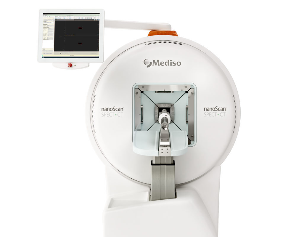
Results from the nanoScan PET/CT and nanoScan® SPECT/CT

Fig1. Na[18F]F PET/CT images in vivo obtained 24 h after coronary artery ligation. After Na[18F]F injection via the tail vein (dose = 18.5 MBq), PET/CT images were acquired using the animal-dedicated scanner (NanoPET/CT, Mediso) under inhalation anesthesia (2–3 % isoflurane in 2–5 l/min of oxygen). CT images were first acquired and then PET images were obtained 30 min after the injection of Na[18F]F. Compared with the control rat (a, b), the rat with MI readily shows strong uptake of Na[18F]F in the infarct (c, d). Panels a and c are transaxial images, and panels b and d are coronal images.

Fig 5. Comparison of [99mTc]MIBI SPECT/CT and Na[18F]F PET/CT images of a rat 1 day after MI. [99mTc]MIBI SPECT/CT and Na[18F]F PET/CT were serially performed in a 2-h interval. Perfusion impairment was readily observed in the apico-lateral area (a, d), where Na[18F]F uptake was positive (b, e). The fusion images of [99mTc]MIBI SPECT and Na[18F]F PET show the close association between the perfusion impairment and Na[18F]F uptake (c, f). Iodinated contrast was not used for the CT acquisitions. Panels a, b, and c are transaxial images, and panels d, e, and f are coronal images.
How can we help you?
Don't hesitate to contact us for technical information or to find out more about our products and services.
Get in touch
