Preclinical evaluation of [11C]GW457427 as a tracer for neutrophil elastase
2022.01.11.
S. Estrada et al., Nuclear Medicine and Biology, 2022
Summary
Inflammatory reactions as primary or secondary phenomena are involved in most of human diseases. The pathological representations of inflammation include many different aspects of tissue reactions such as chronic upregulation of proteolytic enzymes, fibrinogenic cytokines, angiogenic factors, growth factors as well as autoimmune responses. In this process immune active cells, such as leukocytes play a key role.
Neutrophils are the most abundant leukocytes in the circulation. They are part of the innate immune system acting as first line of defense in the immune response and found in many inflammatory diseases, notably in inflammatory bowel disease (IBD) mainly ulcerative colitis (UC) and lung diseases like cystic fibrosis (CF), bronchiectasis (BE) and chronic obstructive pulmonary disease (COPD). Whilst abdominal aortic aneurysm (AAA) is not regarded primarily as an inflammatory disease, involvement of inflammatory cells in the destruction of elastin in the vessel wall is the primary driver of aneurysm formation.
Neutrophils are also known to be involved in the devastating immuno-inflammation found in Acute Respiratory Distress Syndrome (ARDS) and furthermore, it was recently hypothesized that the same mechanism could be responsible in the secondary COVID-19 pulmonary inflammation.
A key event of neutrophil activity is the release of the protease neutrophil elastase (NE) via degranulation of azurophilic granules creating a high local concentration of NE, either bound to the outer cell membrane of the neutrophils or in the form of neutrophil extracellular traps (NETs).
A key purpose of NE is to degrade components of the extracellular matrix, for example elastin, thereby allowing granulocytes and macrophages to traverse tissue and reach sites of inflammation. NE also cleave the pre-formed proteins into pharmacologically active signaling peptides which act as regulators of cellular function promoting chemotaxis of other immune cells to the inflamed tissue. This overactive immune response may exacerbate the patient's condition and worsen the outcome of infection. The proteolytic activity of NE is balanced in vivo by the antiprotease α1-antitrypsin.
Neutrophil elastase has been a target for drug development in pulmonary disease with therapies aiming to protect lung tissue. Several drugs have been investigated in clinical trials, for example Sivelestat® and AZD9668, with mixed results and no conclusive evidence of efficacy although some positive results are reported. In these studies no molecular imaging modality, such as positron emission tomography (PET), was used to investigate the in vivo interaction between drug and NE and it can be hypothesized that the lack of clinical efficacy either was due to too short treatment period or too low dose.
In AAA, elastase activity contributes to elastin degradation and aneurysm formation throughout disease progression. Elastase inhibition has been shown to reduce aneurysm growth in relevant animal models but at present, no medical therapies, established or in development are available that can halt aneurysm expansion in patients. Although functional imaging of AAA patients suggests that inflammation in the aortic wall contributes to its degradation, these results are inconclusive. The concentration of elastase in the azurophilic granules is high (about 5–10 mM) which leads to significant local concentration of the protease upon release, enough to be measured with a selective and specific PET-radioligand. For this purpose, the authors have developed [11C]GW457427 (coined [11C]NES) as a selective and specific PET-tracer for the study of neutrophil elastase in vivo in humans using PET, here validated in vitro in human abdominal aortic aneurysm tissue compared with healthy aorta and in vivo in a lipopolysaccharide (LPS) animal model of lung inflammation compared with control animals. The authors also present a GMP validated production method and toxicology data for [11C]GW457427.
Results from nanoScan PET/MRI 3T and nanoScan SPECT/CT
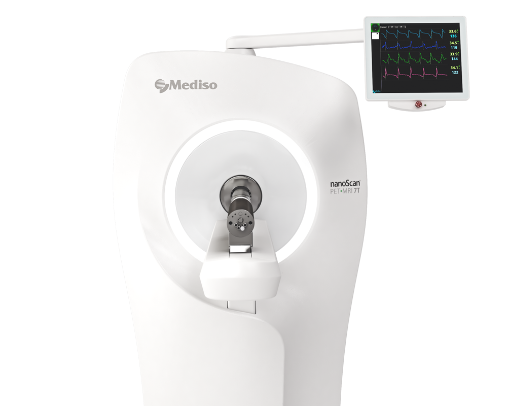
For the imaging studies, [11C]GW457427, 2–5 MBq, in saline, was administered intravenously (i.v.) as a bolus to three groups of BALB/c mice: (i) animals with induced inflammation (n = 8), (baseline group); (ii) animals with induced inflammation (using LPS and Formylmethionyl-leucyl-phenylalanine - fMLP) (n = 6), which also received a co-injection of excess of unlabelled GW457427 (1 mg/kg) (blocked group) and (iii) untreated BALB/c mice (n = 6, control group). In vivo dynamic PET imaging was performed in two animals from each group (n = 6), followed by a CT scan. Mice were positioned in a heated bed of the PET/MRI 3T scanner and a dynamic PET acquisition was initiated immediately before injection of [11C]GW457427. The duration of the PET scan was 40 min, with the thorax in the field of view. After the PET acquisition, the bed was transferred to the Mediso SPECT/CT. A whole-body CT scan was then performed for 8 min. PET images from the Mediso system were reconstructed using a Tera-TomoTM 3D algorithm with 4 iterations and 6 subsets. PET-CT images were fused and analyzed in PMOD 4.0.
Figure 5. shows that the dynamic PET studies were in good agreement with ex vivo data and similarly showed high uptake in the lungs of treated (LPS + fMLP) animals but abolished in the control and the blocked group. A direct comparison between lung-SUV values determined by PET and by ex vivo measurements is not possible, but, assuming a lung density of 0.3 g/mL, SUV values would have been ~6 at the end of the PET acquisition (40 min).
As it is on Figure 7., Dynamic PET also revealed the kinetics of uptake in other organs in mice. For control mice, this was similar to the uptake and clearance from organs in rat, obtained in the ex vivo dosimetry study, and showed rapid wash-out kinetics in the majority of the organs, transient accumulation in liver and kidneys, increased radioactivity in the knee joint and in the intestines suggesting hepatobiliary elimination and specific uptake in red bone marrow, where neutrophils are generated and stored.
- In vivo imaging studies have revealed very low tracer uptake in healthy tissue, apart from bone marrow, with some distribution in the intestines due to hepato-biliary excretion.
- Small animal PET-CT-MR studies in the LPS pulmonary inflammation model in mice as described showed high uptake in lungs which could be blocked by co-injection of GW457427.
- The specificity in binding to elastase was confirmed in vitro, using the two structurally different selective elastase inhibitors, Sivelestat® and GW311616. Hence, [11C]GW457427 is a very promising PET biomarker for NE and currently, it is being investigated in a First-In-man study.
How can we help you?
Don't hesitate to contact us for technical information or to find out more about our products and services.
Get in touch
