A tool for nuclear imaging of the SARS-CoV-2 entry receptor: molecular model and preclinical development of ACE2-selective radiopeptides
2023.04.19.
Darja Beyer et al., 2023, EJNMMI Research
Summary
Covid-19, an infectious disease caused by the SARS-CoV-2 virus, was declared a pandemic by the World Health Organization in March 2020 and the disease has become a significant global health threat to society. The numerous virus variants have contributed to variable pathological manifestations, symptom severity and long-term effects of Covid-19. SARS-CoV-2 uses the angiotensin converting enzyme-2 (ACE2) as an entry receptor for host cell infection. ACE2 was discovered in the year 2000 and identified as an essential enzyme for cardiovascular regulation processes. The catalytic domain of ACE2 shares 42% sequence identity with its homologue, the angiotensin-converting enzyme (ACE); however, these enzymes differ in terms of substrate selectivity and physiological functions. Importantly, ACE2 does not bind ACE inhibitors such as captopril or lisinopril, which are part of a frequently used class of drugs to treat hypertension. ACE2 has an antihypertensive and cardioprotective role and acts as counter regulator of ACE in the renin–angiotensin–aldosterone system (RAAS). ACE2 was found to be highly expressed in the cardiovascular system, lungs, kidneys, intestines and testis. Not surprisingly, several of these tissues were identified as critical sites in Covid-19 pathological manifestations.
The complex regulation mechanisms of ACE2 expression, including dynamic changes after SARS-CoV-2 infection, may be one of several reasons why Covid-19 displays such large variability of manifestations and progression ranging from asymptomatic to severe cases with fatal outcomes. Some studies propose a positive correlation between ACE2 expression and disease severity as a result of enhanced viral propagation, and argue that lower ACE2 levels in children protects them from severe SARS-CoV-2 infections. Other studies suggest a poor prognosis of elderly as a result of low ACE2 expression. The contradictory statements with regard to age-related ACE2 expression may originate from strong inter-individual variation, cell-type-dependent maximum age of ACE2 degradation and the fact that mRNA and protein levels do not exactly correspond. There is also the theory that the degradation of ACE2 through infection with SARS-CoV-2 and, thus, the lack in ability to counterbalance the action of ACE in the RAAS may lead to a pro-inflammatory state and, therefore, an increased risk of a severe disease progression.
The hypothesis that ACE2 expression levels and dynamics affects Covid-19 severity is further supported by the observation that medical conditions that can affect the ACE2 expression levels such as hypertension, diabetes and obesity, but also genetic predisposition and certain medications, are known risk factors for severe disease phenotypes of Covid-19.
A better understanding of the dynamics of ACE2 expression as a means to predict the susceptibility to infection and disease progression would possibly enable identifying patients at risk and allow the development of more individualized treatment strategies. While attempts have been made to determine ACE2 in various tissues at the mRNA and protein levels, noninvasive imaging would also enable to determine the expression dynamics directly in patients.
Among the imaging techniques used in the clinic, single photon emission computed tomography (SPECT) is still more often employed than positron emission tomography (PET). The enhanced image resolution, the increased sensitivity and the option for accurate quantification present, however, relevant advantages of PET, which would, therefore, be preferred for molecular imaging of (patho)physiological processes of many medical situations, including cardiovascular diseases. Preclinical attempts to develop a PET imaging agent to visualize ACE2 were initially made by labeling the receptor-binding domain of the viral spike protein with iodine-124. Other research groups used the disulfide-bridged DX600, initially developed as an ACE2-specific inhibitor, for derivatization with a DOTA or NODAGA chelator to enable coordination of gallium-68 as a PET radionuclide. A NOTA-derivatized DX600 peptide, referred to as DX600-BCH, was developed for coordination of aluminium-fluoride-18 and is currently being used in a clinical trial to test the possibility of noninvasive mapping of ACE2 (NCT04542863).
The authors' aim was to perform a more in-depth investigation of DX600-based radiopeptides for noninvasive PET imaging of ACE2. With the aid of molecular modeling, three peptides with a DOTA, a NODAGA or a HBED-CC chelator, respectively, were designed. The three DX600 peptides were evaluated after labeling with gallium-67 (T1/2 = 3.26 d), as a surrogate radioisotope for gallium-68 (T1/2 = 68 min), due to its more convenient half-life and because it can be used for SPECT. The ACE2 selectivity of the DX600-based radiopeptides was assessed by the use of ACE2- and ACE-transfected human embryonic kidney (HEK) cells and respective xenograft mouse models. An ACE-specific radiopeptide based on the structure of the bradykinin-potentiating peptide-9a (BPP9a) was synthesized and derivatized for metal chelation for the purpose of validating the in vitro and in vivo models.
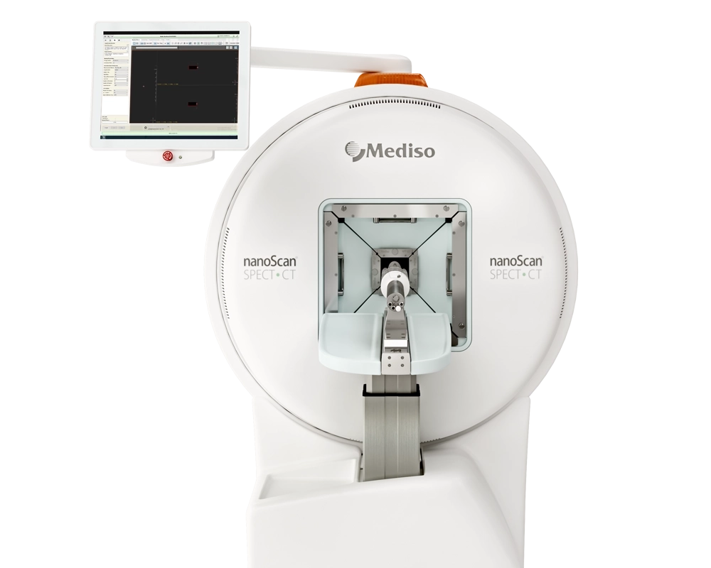
Results from the nanoScan SPECT/CT
SPECT/CT imaging was performed using a four-head, multiplexing, multipinhole small-animal SPECT camera (NanoSPECT/CT™). Each head was outfitted with a tungsten-based aperture of nine 1.4 mm-diameter pinholes and a thickness of 10 mm. CT scans of 7.5 min duration were followed by SPECT scans of ~ 50 min performed 1 h, 3 h and 24 h after injection of the respective radiopeptide (10 MBq, 0.5 nmol, 100 μL), diluted in NaCl 0.9% containing 0.05% BSA. During the scan, mice were anesthetized with a mixture of isoflurane and oxygen. The images were acquired using Nucline software. The real-time CT reconstruction used a cone-beam-filtered backprojection. The reconstruction of SPECT data was performed with the HiSPECT software using γ-energies of 93.20 keV (± 10%), 184.60 keV (± 10%) and 300.00 keV (± 10%) for gallium-67. All images were prepared using the VivoQuant post-processing software. A Gauss post-reconstruction filter (full width at half maximum, 1 mm) was applied, and the scale of activity for gallium-67 was set as indicated on the SPECT/CT images. In order to estimate the wash-out of the DX600-based radiopeptides from the HEK-ACE2 xenografts and kidneys over time, the absolute amount of activity was determined in these tissues using the VivoQuant quantification tool. The accumulated activity (non-decay-corrected value in MBq) in the xenografts and kidneys, respectively, obtained at the 1 h p.i. timepoint was set as 100%. The percentage of activity retained in these tissues was determined at 3 h and 24 h p.i. based on the absolute activity determined at these timepoints.
SPECT/CT imaging studies of mice conducted at 1 h and 3 h after injection of the DX600-based radiopeptides revealed the activity accumulation in ACE2-expressing xenografts but not in those that expressed ACE. The radiopeptides were well retained in the HEK-ACE2 xenografts over time showing still about 92–95% and about 47–58% of the activity in the xenografts after 3 h and 24 h, respectively, compared to the 1 h p.i.-timepoint. All three radiopeptides showed retention in the kidneys as a result of renal excretion. This was particularly prominent after injection of [67Ga]Ga-DOTA-DX600 and [67Ga]Ga-NODAGA-DX600 (Fig. 7A/B), for which ~ 85% of the measured activity at the 1 h p.i.-timepoint was retained after 3 h and still about 35–45% after 24 h. In the case of [67Ga]Ga-HBED-CC-DX600, kidney clearance was much faster (Fig. 7C), demonstrated by only 30% of the activity left in the kidneys after 3 h relative to the amount measured at the 1 h p.i.-timepoint. After 24 h, [67Ga]Ga-HBED-CC-DX600 was entirely excreted from the kidneys (< 1% of the activity measured at the 1 h p.i.-timepoint).
- The authors' data identified [67Ga]Ga-HBED-CC-DX600 as a promising nuclear imaging agent to investigate the in vivo expression profile and dynamics of ACE2 for a better understanding of Covid-19 pathology.
- The biodistribution of [67Ga]Ga-HBED-CC-DX600 outperformed that of other DX600-based radiopeptides with DOTA or NODAGA chelators. Importantly, the HBED-CC chelator enabled 67Ga-labeling at high molar activity, which would be essential to obtain images with high signal-to-background contrast to detect (patho)physiological ACE2 expression levels in patients.
Hogyan segíthetünk Önnek?
További termékinformációkért, vagy támogatásért keresse szakértőinket!
Vegye fel a kapcsolatot
